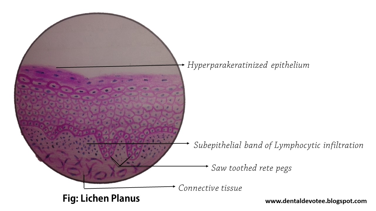
SEARCH:

Modified Essay Questions, MEQs in General Surgery - Wound

WOUND 

1. A 9 yr old boy was hit by a vehicle while going to school a few hours back. He was carried to the emergency in a stable state with wound on right calf.         (15 marks) 
a. Define wound.
Wound is the discontinuity or break in the surface epithelium

b. Describe the local pathophysiology of wounds.
- Stage of hemostasis - Stage of  inflammation - stage of granulation - stage of maturation

On examination, 15 cm x 6 cm wound defect was noticed over the postero-lateral aspect of rt leg which had ragged, unhealthy margins and there was skin loss as well. 

2. Classify open wounds.
i. Incised wound: superficial wound with a sharp edge and caused by sharp objects less contaminated
ii. Lacerated wounds: caused by blunt objects or RTA. Ragged unhealthy edges,  crushing or tissue may be present
iii. Penetrating wounds: caused by sharp objects, depth of wound greater than length. Internal organs, blood vessels, nerves might be damaged
iv. Crushed wounds: Caused by blunt trauma eg RTAs. Might cause severe hemorrhage,
more prone for gangrene, tetanus etc.

3. Define Abrasion and Laceration.
- Abrasion: Scraping or peeling off of the epidermis with the exposure of the dermis.
- Laceration: Laceration is the tearing or splitting of skin, mucous membranes, muscles or internal organs caused by either a shearing or a crushing force, and produced by application of a blunt force to a broad area of the body.

Parents of the boy are anxious. They want to know how you will manage their son.

4. Outline the treatment plan.
 i. Resuscitation if the patient unstable. Not required here as the patient is stable
ii. Anagesics if the patient is in pain -WHO step ladder. NSAIDs followed by opoids
iii. Cleaning of wound
iv. Exploration and diagnosis
v. Debridement
vi. Repair of Structures, splinting of fractures
vii. Replacement of lost tissue where indicated
viii. Skin cover if required
ix. Skin closure without tension
x. Tetanus Toxoid, Antibiotics (broad spectrum including anaerobic coverage)

5. What is the most important point that you will consider while doing a primary closure:
- presence of infection (if present it cannot be done)

6. Define Healing by primary intension.
- It is the healing of clean incised wounds such as surgical incision by re-epithelialization across the wound producing a neat thin scar.

7. Somehow you have managed to close the wound by primary closure but the next day you findthat the wound edges have turned black. What will you do next?
- Open the sutures, take swabs for cultures debride necrotic tissue and leave the wound open. Continue antibiotics.

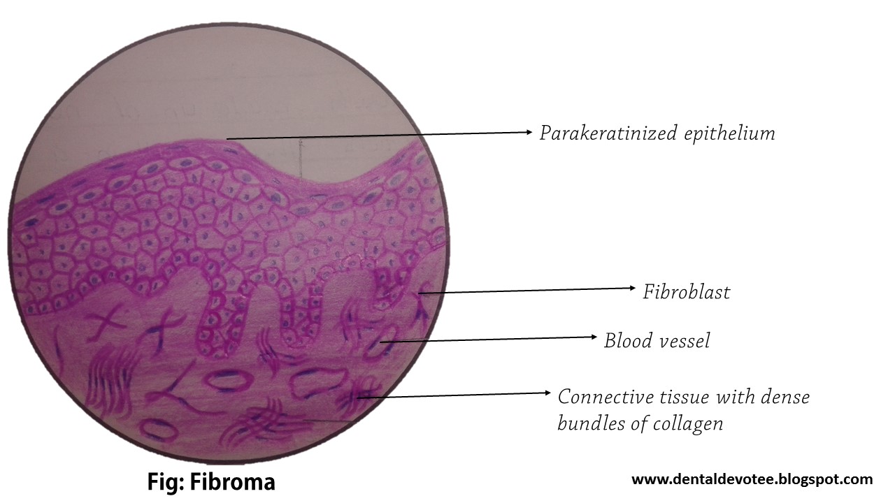
8. Define Granulation tissue. 
- It is a soft, pink, granular tissue that is formed during wound healing. It consists of small new blood vessels and proliferation of fibroblasts.

9. What is healing by secondary intention?
- It is the type of healing that takes place in wounds with unopposed edges where there is granulation, contraction, and epithelialization.

10. What is the next best alternative if one cannot close the wound primarily? 
- Repair by delayed primary repair: Wound initially left open and edges later apposed when the healing conditions are favorable. 

Important Definitions in Oral Pathology

Neoplasm
A neoplasm, as defined by Willis, is 'an abnormal mass of tissue the growth of which exceeds and is uncoordinated with that of the normal tissues and persists in the same excessive manner after the cessation of the stimuli which evoked the change.'

Premalignant / Precancerous lesion
A morphologically altered tissue in which cancer is more likely to occur than in its apparently normal counterpart.  - WHO 1973

Premalignant / Precancerous condition
A generalized state associated with a significantly increased risk of cancer. - WHO 1973

Potentially malignant Oral Diseases
The term Potentially Malignant Disorders was recommended to refer to precancer as it conveys that not all disorders described under this term may transform into cancer.  - Warnakulasuriya et al 2007

Leukoplakia
White patch or plaque that cannot be characterized clinically or pathologically as any other disease. - WHO 1973

Leukoplakia should be used to recognize white plaques of questionable risk having excluded (other) known diseases or disorders that carry no increased risk of cancer.

It is to be noted that a lesion of leukoplakia is non-scrappable.

Erythroplakia
A fiery red patch that cannot be characterized clinically or pathologically as any other definable disease.  - WHO 1973

Oral Submucous Fibrosis
It is an insidious chronic disease affecting any part of the oral cavity and sometimes the pharynx. Although occasionally preceded by and/or associated with vesicle formation, it is always associated with a juxta- epithelial inflammatory reaction followed by a fibro-elastic change of the lamina propria with epithelial atrophy leading to stiffness of the oral mucosa and causing trismus and inability to eat.
- Pindborg and Associates

Cyst
A cyst is defined as ' a pathological cavity having fluid, semifluid or gaseous contents and which is not created by the accumulation of pus'. Most cysts, but not all, are lined by epithelium. - Kramer, 1974

Oncology: the study of neoplasms


Neoplasm: an uncontrolled new growth of tissue

Tumor: a localized swelling, may or may not be a true neoplasm

Hyperplasia: An increase in the size of a tissue or organ due to an increase in the number of component cells

Hypertrophy: an increase in the size of a tissue or organ due to an increase in the size of component cells

Cancer: a general term for all malignant neoplasms

Carcinoma: a malignant epithelial neoplasm

Sarcoma: a malignant mesenchymal (connective tissue) neoplasm

Hamartoma: a developmental defect characterized by an overgrowth of tissues normal to the organ in which it arises

Teratoma: a neoplasm composed of multiple tissues foreign to the organ in which it arises; may be benign or malignant


Dental Amalgam : Uses, Classification, Advantages and Disadvantages

An amalgam is defined as a special type of alloy in which mercury is one of the components. Dental amalgam is the most widely used filling material for posterior teeth. As one of the oldest restorative material, dental amalgam has evolved over time and has become successful to stand the test of time.
The quantity of mercury has been reduced over time and newer and newer components are incorporated in the alloy powder to reduce the corrosion of dental amalgam in the oral cavity.

HISTORY
Louis Regnart added mercury to the D’Arcet’s Mineral Cement mixture, which was widely used in France then, lowering the temperature required to boil the mixture significantly, and for this became known as the ‘Father of Amalgam’. Early amalgam was made by mixing mercury with the filings of silver coins. Crawcour brothers (from France) introduced the amalgam in the United States of America in 1833 which used to have significant expansion which even caused tooth fractures in some restorations. However, this challenge was overcome when in 1895, GV Black developed a
formula (67% silver, 27% tin, 5% copper, 1% zinc) for modern amalgam alloy. Black’s formula overcame the expansion problems of the existing amalgam formulations.

APPLICATIONS
1. As a permanent filling material for
— Class I and class II cavities, and
— Class V cavities where aesthetics is not important.

2. In combination with retentive pins to restore a crown

3. For making dies

4. In retrograde root canal fillings

5. As a core material.

CLASSIFICATION OF AMALGAM ALLOYS
BASED ON COPPER CONTENT
Low copper alloys: Contain less than 6% copper (conventional alloys)
High copper alloys: Contain between 13-30% copper.

The high copper alloys are further classified as:

  • Admixed or dispersion or blended alloys.
  • Single composition or unicomposition alloys.

BASED ON ZINC CONTENT
Zinc-containing alloys: Contain more than 0.01% zinc
Zinc-free alloys: Contain less than 0.01% zinc

BASED ON SHAPE OF THE ALLOY PARTICLE
Lathe cut alloys (irregular shape)
Spherical alloys
Spheroidal alloys

BASED ON NUMBER OF ALLOYED METALS
Binary alloys, e.g., silver-tin
Ternary alloys, e.g., silver-tin-copper
Quaternary alloys, e.g., silver-tin-copper-indium.

BASED ON SIZE OF ALLOY
Micro-cut
Macro-cut

ADVANTAGES AND DISADVANTAGES OF AMALGAM  RESTORATIONS
Advantages
1. Reasonably easy to insert.
2. Not overly technique sensitive.
3. Maintains anatomic form well.
4. Has adequate resistance to fracture.
5. After a period of time prevents marginal leakage.

6. Have reasonably long service life.
7. Cheaper than other alternative posterior restorative material like cast gold alloys.

Disadvantages
1. The color does not match tooth structure.
2. They are more brittle and can fracture if incorrectly placed.
3. They are subject to corrosion and galvanic action.
4. They eventually show marginal breakdown.
5. They do not bond to tooth structure.
6. A risk of mercury toxicity.

Oral Pathology Histopathological Diagrams - All in one - Histology made easy


This post contains 33 sorted diagrams of histopathological pictures of pathologies related to oral and maxillofacial regions. From benign to malignant neoplasms of oral cavity, salivary gland tumors, cyst and tumors of jaws and oral cavity, to lesions affecting nerves and muscles and soft tissues, it contains all you need to have a good command in oral pathology. 

These diagrams were drawn with Hematoxylin and Eosin colored Pencils by Raman Dhungel during his Bachelor of Dental Surgery course on third year at B.P. Koirala Institute of Health Sciences, BPKIHS Dharan.  The slides included in this video are: Fibroma, FIbrosarcoma, Papilloma, Verrucous carcinoma, Mild dysplasia, Carcinoma in situ, Squamous cell carcinoma (well differentiated), Neurilemmoma, Neurofibroma, Oral submucous fibrosis, cavernous hemangioma, Lipoma, Lichen planus, malignant melanoma, osteosarcoma, pleomorphic adenoma, warthin's tumor, Adenoid cystic carcinoma, Mucoepidermoid carcinoma, Calcifying epithelial odontogenic tumor, Adenomatoid odontogenic tumor, Keratocystic odontogenic tumor, Dentigerous cyst, Radicular cyst, Plexiform ameloblastoma, Follicular ameloblastoma, Central giant cell granuloma, Peripheral giant cell granuloma, Fibrous dysplasia, Paget's disease of bone, Pemphigus, Pemphigoid (Bullous pemphigoid), and pyogenic granuloma.

Much time and effort was needed to produce this post. We would be gratified to know that if this postbecomes of some help to you. Please share with your friends and students too. 


Behcet's Syndrome

Behçet’s syndrome is a multisystem disease that predominantly affects young males and is
characterized by multiple superficial, painful “aphthous-like ulcers” in the oral cavity.
However to fulfill the criteria of being Behçet’s syndrome, clinically there should be a presence of an aphthous-like ulcer in the oral cavity along with at least two of the following lesions e.g. skin lesion, eye lesion or genital lesion, etc.

ETIOLOGY
Etiology of Behcet’s syndrome is unknown; however, the disease is believed to be caused by
some immunologic abnormality.

CLINICAL FEATURES
Oral lesions: Aphthous-like ulceration in the oral cavity.

Skin lesions: Erythematous macular, papular, vesicular or pustular lesions in the skin; thrombophlebitis may also sometimes develop.

Eye lesions: Ocular lesions in Behçet’s syndrome include uveitis, conjunctivitis, photophobia and retinitis, etc.

Genital lesions: Ulceration in the genitalia, which looks similar to those of the oral cavity.

Other lesions: Behcet’s syndrome sometimes presents some additional features like neural,
vascular, articular, renal or gastrointestinal lesions of various kinds.

HISTOPATHOLOGY
Microscopically the lesions produce similar feature to what is found in a minor aphthous ulcer. However, there can be some additional features like severe vasculitis and vascular damage, etc.

TREATMENT
Behçet’s syndrome is treated by systemic steroid therapy.

Pyogenic Granuloma

Pyogenic granuloma represents an over-exuberant tissue reaction to some known stimuli or injuries. The term pyogenic granuloma is somewhat a misnomer since the condition is not associated with pus formation.

CLINICAL FEATURES
Age: occurs at an early age.
Sex: seen more frequently in females.
Site: mostly occurs in relation to the gingiva, however on rare occasions, other mucosal sites may be involved.

PRESENTATION
• The lesion appears as a small, pedunculated or sessile, painless, soft, lobulated growth on the gingiva

• Labial surface of the gingiva is more frequently affected than the lingual surface.

• The lesion is often ulcerated and bleeds profusely, either upon provocation or spontaneously.

• The ulcerated area of the lesion is often covered by a yellow fibrinous membrane.

• The rate of growth of the lesion is very rapid and its maximum size could be up to 1 cm in
diameter

•Untreated lesion of pyogenic granuloma undergoes fibrosis due to decreased vascularity and in such cases it appears small, firm with little tendency to bleed. This lesion is called “fibroepithelial polyp”

• Similar lesions appearing on the gingival tissue of pregnant women are known as “pregnancy tumor”.

HISTOPATHOLOGY 
• Histologically, the lesion is composed of lobular masses of hyperplastic granulation tissue, containing multiple proliferating fibroblasts, many blood capillaries and a variable number of chronic inflammatory cells.

• The lesion is a vascular one and it occurs due to the proliferation of the endothelial cells.

• The overlying epithelium is thin and ulcerated, and in most of the cases the underlying
connective tissue shows intercellular edema.

• Areas of hemorrhage and hemosiderin pigmentation are often seen within the connective tissue stroma.

TREATMENT
Pyogenic granuloma is treated by surgical excision.

Oral Biology Past Question - 2nd Year BDS BPKIHS March 2016

Time: 2 Hours                             Total Marks: 125
1. Mention five points of difference between cellular and acellular cementum.    - 5

2. Mention five points of difference between primary and permanent pulp tissues.    - 5

3. Define Crest of Curvature. Describe the Crest of Curvature of a maxillary canine from all four aspects with a neat labeled diagram.    - 2+4 = 6

4. Describe six points of difference between primary and permanent human dentition.      - 6

5. Describe the labial aspect of the permanent mandibular right lateral incisor.      - 6

6. List and discuss the functions of dental pulp.    - 6

7. Mention the different types of the cementoenamel junction and discuss the formation of any one.  - 6

8. Discuss the phenomenon of formation of Hunter-Schreger band.      - 6

9. Describe late bell stage of amelogenesis with suitable diagram.       - 4+3 = 7

10. List the transitory structures formed during the development of teeth and describe any one.     - 7

11. Discuss the development of periodontal ligament fibers.     - 7

12. Discuss the formation of the root of the permanent mandibular central incisor.   - 7

13. Describe the process of modulation in amelogenesis with suitable diagrams.      - 7

14. Discuss the keyhole pattern of enamel.    - 7

15. Discuss reciprocal induction in context to the development of teeth.     -7

16.  Discuss in detail the anatomy of the permanent maxillary right central incisor.    -10


17. WRITE SHORT NOTES ON: (3*5 = 15)
a. Perikymata
b. Prismless enamel
c. Intermediate plexus
d. Neural crest cells
e. Plexus of Rashkow

18. Define the following. (1*5 = 5)
a. Fossa
b. Ridge
c. Lobe
d. Cingulum
e. Point angle