SEARCH:

MCQs on Dental Materials : Amalgams Part 1


# The term "trituration" means :
A. Lysing amalgam alloy
B. Mixing of amalgam alloy and mercury
C. Removal of excess of mercury
D. None of the above

# Dynamic creep is the:
A. Continuing alloying between Silver-Tin alloy and mercury during the life of the restoration
B. Deformation of set amalgam during function
C. Process whereby alloy is wetted by mercury
D. Spread of amalgam during packing

Tooth Sensitivity / Dentin Hypersensitivity

Introduction
The Canadian advisory board on dentin hypersensitivity ( 2003 ) defined Dentin hypersensitivity (Tooth sensitivity) as “a short, sharp pain arising from exposed dentin in response to stimuli typically thermal, evaporative, tactile, osmotic or chemical and which cannot be ascribed to any other form of dental defect or disease.”  It has been described as the “common cold of dentistry” by some and
“toothbrush disease” by others when it occurs in the presence of gingival recession (Pashley et al. 2008 ).

Prevalence and Distribution
The condition is mostly prevalent among the young population in the 3rd and 4th decades. The prevalence may shift in the future to a younger age group because of the increase in acidic food/drink intake and the influence of greater oral hygiene awareness and measures (Chabanski et al. 1997; Clayton et al. 2002 ). Various intraoral locations can be affected with Dentin hypersensitivity. Sites of predilection in descending order are canines and first premolars, incisors and second premolars, and molars (Dababneh et al. 1999 ). The buccal surfaces are mostly affected, followed by labial, occlusal, distal, and lingual. Incisal and palatal surfaces are the least affected (Splieth and Tachou 2012; Amarasena et al. 2011 ).

Mechanism
Brännström’s ( 1962, 1992 ) hydrodynamic theory of dentin sensitivity proposed that hydrodynamic
stimuli (hot or cold, tactile, evaporative or osmotic) caused sudden minute shifts of dentinal fluid that activate pulpal mechanoreceptors to cause sharp, well-localized tooth pain, thought to be due to A-delta sensory nerves (Narhi et al., 1992 ).

Causes and Predisposing Factors
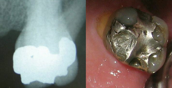
The most important factor to be present for tooth sensitivity is the loss of tooth structure and the exposure of dentin to the oral cavity. The dentin may be exposed to the oral cavity by either the loss of enamel or cementum and overlying periodontal attachment apparatus or loss of both at the same time. Following the exposure, the patent dentinal tubules remain wide open and thus are predisposed to any stimulus, called the phase of “lesion initiation.” However, not all exposed dentin is sensitive (Rimondini et al. 1995 ).

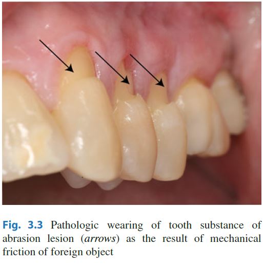
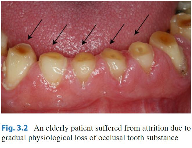
The loss of enamel may be either due to attrition, erosion, abrasion, abfraction or abrasion due to tooth brushing (most commonly cervical abrasion). Similarly, loss of cementum due to various causes and periodontal attachment loss may cause exposure of dentinal tubules. Some medical conditions like Bulimia Nervosa, Gastroesophageal reflux disease, Chronic alcoholism, Salivary hypofunction, etc. may also cause dentin hypersensitivity.


Diagnosis
Diagnosis of dentin hypersensitivity can be made by applying any of the mechanical, chemical, thermal or electrical stimuli. Electronic pulp testers (EPT), mechanical pressure stimulators, air jet stimulator, ethyl chloride, thermoelectric device, cold water testing, ice testing, etc. can be used for challenging the affected tooth.

Treatment 
Two treatment approaches are mainly practiced in the treatment of dentin hypersensitivity. They are:

  •  Use of dentin blocking agents that occlude patent (open) tubules (fluoride, strontium salts, oxalate, calcium phosphate, restorative materials, etc.), and
  • Nerve desensitization agents that reduce intradental nerve excitability (e.g. potassium ions, guanethidine) in order to prevent a response from intra dental nerves to the stimulus- evoked fluid movements within the dentin tubules


 Application of these successfully tested products may either involve ‘in-office’ procedures by a clinician using a restorative approach (for example, restorative materials in the form of dentin bonding agents, glass ionomer cements (GIC), and periodontal surgical techniques) or by a clinician recommending an over-the-counter (OTC) approach (involving toothpastes, gels, mouthwashes).

Furthermore, the causative agent has to be identified and avoided as much as possible and the patient should be educated about the proper brushing technique to prevent mechanical abrasion due to toothbrushing in the future.


References:
1. Canadian Advisory Board on Dentin Hypersensitivity (2003) Consensus-based recommendations for the diagnosis and management of dentin hypersensitivity. J Can Dent Assoc 69(4):221–226

2. Pashley DH, Tay FR, Haywood VB, Collins MA, Drisko CL (2008) Consensus-based recommendations for the diagnosis and management of dentin hypersensitivity. Inside Dentistry 4(9 (Special Issue)):1–35

3. Chabanski MB, Gillam DG, Bulman JS, Newman HN (1997) Clinical evaluation of cervical dentine sensitivity in a population of patients referred to a specialist periodontology department: a pilot study. J Oral Rehabil 24(9):666–672

4. Clayton DR, McCarthy D, Gillam DG (2002) A study of the prevalence and distribution of dentine sensitivity in a population of 17-58-year-old serving personnel on an RAF base in the Midlands. J Oral Rehabil 29(1):14–23, 805 [pii]

5. Dababneh RH, Khouri AT, Addy M (1999) Dentine hypersensitivity – an enigma? A review of terminology, mechanisms, aetiology and management. Br Dent J 187(11):606–611; discussion 603. doi: 10.1038/sj.bdj.4800345a , 4800345a [pii]

6. Brännström M (1992) Etiology of dentin hypersensitivity. Proc Finn Dent Soc 88(Suppl 1):7–13

7. Brännström M (1962) The elicitation of pain in human dentine and pulp by chemical stimuli. Arch Oral Biol 7:59–62

8. Närhi M, Jyvasjarv E, Vitanen A, Huopaniemi T, Ngassapa D, Hirvonen T (1992) Role of intra dental A and C type nerve fibers in dental pain mechanisms. Proc Finn Dent Soc 8(Suppl 1):507–516

9. Dababneh RH, Khouri AT, Addy M (1999) Dentine hypersensitivity – an enigma? A review of terminology, mechanisms, aetiology and management. Br Dent J 187(11):606–611. doi: 10.1038/sj.bdj.4800345a ; discussion 603

10. Clinician’s Guide to the Diagnosis and Management of Tooth Sensitivity ; Sahar Taha, Brian H. Clarkson ISBN 978-3-642-45163-8 ISBN 978-3-642-45164-5 (eBook) DOI 10.1007/978-3-642-45164-5

11. Rimondini L, Baroni C, Carrassi A (1995) Ultrastructure of hypersensitive and non-sensitive dentine. A study on replica models. J Clin Periodontol 22(12):899–902

12. Ling TYY, Gillam DG (1996) The effectiveness of desensitizing agents for the treatment of cervical dentine sensitivity (CDS) – a review. Periodontal Abstr 44(1):5–12

13. Orchardson R, Gillam D (2006) Managing dentin hypersensitivity. J Am Dent Assoc 137(7):990–998

Download these Textbooks of Dentistry as soon as possible

These are the important textbooks in Dentistry.

Oral Anatomy and Histology
1. Wheeler's Dental Anatomy, Physiology, and Occlusion    - Stanley J. Nelson
2. Woelfel's Dental Anatomy   - Rickne C. Scheid, Gabriella Weiss
3. Ten Cate's Oral Histology: Development, Structure, and Function    - Antonio Nanci
4. Orban's Oral Histology and Embryology  - G.S. Kumar
5. Oral Anatomy, Histology, and Embryology  - B.K.B. Berkovitz, G.R. Holland & B.J. Moxham

Oral Medicine and Radiology
1. Burket's Oral Medicine  - Michael Glick
2. Oral Radiology: Principles and Interpretation  - Stuart C. White, Michael J. Pharaoh

Oral and Maxillofacial Surgery
1. Peterson's Principles of Oral and Maxillofacial Surgery
2. An Introduction to Oral and Maxillofacial Surgery  - David A. Mitchell
3. Handbook of Local Anesthesia   - Stanley F. Malamed

Pediatric Dentistry / Pedodontics
1. Dentistry for the Child and Adolescent     - Ralph E. McDonald, David R. Avery
2. Principles and Practice of Pedodontics      - Arathi Rao

Orthodontics
1. Contemporary Orthodontics   - William R. Proffit
2. Orthodontics The art and Science - S.I. Bhalajhi
3. Orthodontics - Current Principles and Techniques - Graber, Vanarsdall ang Vig

Public  Health Dentistry
1. Jong's Community Dental Health   - George M. Gluck & Warren M. Morganstein
2. Dentistry, Dental Practice, and the Community  - Brian A. Burt, Stephen A. Eklund
3. Park's Textbook of Preventive and Social Medicine  - K. Park

Prosthodontics
1. Prosthodontic Treatment for Edentulous Patients     -    Zarb, Hobkirk, Eckert and Jacob
2. McCracken' Removable Partial Prosthodontics    - Alan B. Carr & David T. Brown


Periodontology
1. Carranza's Clinical Periodontology  - Newman, Takei, Klokkevold & Carranza
2. Clinical Periodontology and Implant Dentistry   - Niklaus P. Lang & Jan Lindhe

Operative Dentistry
1. Sturdevant's Art and Science of Operative Dentistry  - Harald O. Heymann, Edward J. Swift & André V. Ritter
2. Pickard's Manual of Operative Dentistry  - Avijit Banerjee & Timothy H. Watson

Oral Pathology
1. Shafer's Textbook of Oral Pathology  -  R. Rajendran & B. Sivapathasundharam
2. Oral and maxillofacial Pathology  - Neville, Damm, Allen, and Bouquot
3. Oral Pathology : Clinical pathologic Correlation  - Regezi, Sciubba & Jordan

BPKIHS Past Question : Endocrinology and Reproductive System - 2nd year MBBS

B.P. Koirala Institute of Health Sciences, Dharan
2nd Year MBBS
Unit - 4
Internal Assessment, January 2000

Paper - IIA
(Endocrines & Reproduction)

Time: 2 hour                                                                                                                                Maximum Marks: 125

Short Answer Questions

Please answer each section in a separate answer book.

                                             Section 'A'
Anatomy
1.                Draw a neatly labeled histological diagram of a mature ovarian follicle (Grafian follicle).
Explain how theca externa is formed.                                                                                    3+1

2.            Define deep perineal pouch. Enlist six components of the deep perineal pouch.               2+3

3.            Explain in brief why incisions into the breast are usually made radially ?                             3

4.            Explain in brief the embryological basis of  thyroglossal cyst.                                              3

Acrylic Teeth Vs Porcelain Teeth

Property Acrylic Teeth Porcelain Teeth
Abrasion Resistance LowHigh
Adjustability Easy Difficult
Bonding ChemicalMechanical
Staining Easily stained Does not stain
Percolation Absent when acrylic denture base is usedPresent when acrylic denture base is used
Clicking Sound Absent Present
Ease of Fabrication EasyDifficult
Ease of rebasing Difficult to remove acrylic teeth Esay to remove porcelain teeth
Trauma to dental bearing area LessMore

Anatomical Landmarks in Maxilla and Mandible for Complete Denture Fabrication

Anatomical Landmarks in Maxilla and Mandible for Complete Denture Fabrication

MAXILLA
Primary Stress bearing area Residual alveolar ridge
Secondary stress bearing area
  • Rugae or anterior hard palate
  • Maxillary Tuberosity
Tertiary stress bearing area and secondary retentive area Posterolateral part of the hard palate
Relieving areas
  • Incisive papilla
  • Mid palatine raphe
  • Cuspid eminence
  • Fovea palatinea
Primary retentive area Posterior palatal seal area

MANDIBLE
Primary stress bearing area Buccal shelf area
Secondary stress bearing area Slopes of edentulous ridges
Primary retentive and primary peripheral seal area Retromolar pad
Secondary peripheral seal area Anterior lingual border
Relief areas
  • Crest of the residual ridge
  • Mental foramen
  • Mylohyoid ridge

Classification of Composites and their Properties

Type Properties
Conventional Composite
  • Contain filler particle size of 8-12 micrometers
  • It is the composite with largest filler size
  • High strength and hardness
  • Less water soprtion and Coefficient of thermal expansion
  • Polishing is difficult and results in rough surface that tends to retain stains
Micro filled Composite
  • Contain smallest filler particles of size 0.04 - 0.4 micrometers
  • Colloidal silica is used as a filler
  • It has lowest filler content of 50% weight
  • Lowest strength and hardness
  • Highest thermal expansion coefficient and water sorption
  • Excellent esthetics due to the increased smoothness
Small Particle
  • Contain filler particles of size 1 - 5 micrometers
  • It has good surface smoothness like microfilled composites and improved physical properties like conventional composite
  • It has the highest compressive strength of all composites
Hybrid Composite
  • It has filler particle size of 0.6 - 1 micrometer
  • They have smooth finish and better esthetics than small particle but yet have similar physical properties.