SEARCH:

Periapical granuloma vs Periapical cemental Dysplasia

# Difference between periapical granuloma and periapical cemental dysplasia:
A. Vitality
B. Histologically
C. Radio graphically
D. Culture media


The correct answer is A. Vitality

Periapical granuloma
The tooth in Periapical granuloma is non vital and may be slightly tender to percussion but otherwise symptoms may be minimal.

- Periapical cemento-osseous dysplasia (osseous dysplasia; cemental dysplasia; cementomas).

→ Periapical cemento-osseous dysplasia predominantly involves the periapical region of the anterior mandible.

→ Solitary lesions may occur but multiple foci are present more frequently.

There is a marked predilection for female patients (ranging from 10: 1 to 14: 1) and approximately 70 % of cases affect blacks.

→ Most patients are diagnosed initially between the ages of 30 and 50, with the diagnosis almost never made in individuals under the age of 20 years. 

Teeth associated with the lesions are almost invariably vital and seldom have restorations.

→ Periapical cemento-osseous dysplasia is an asymptomatic condition that is discovered when radiographs are taken for other purposes.

→ Early lesions appear as circumscribed areas of radiolucency involving the apical area of a tooth. At this stage, the lesion cannot be differentiated radiographically from a periapical granuloma or periapical cyst.

With time adjacent lesions often fuse to form a linear pattern of radiolucency that envelopes the apices of several teeth.

Diabetes mellitus symptoms

# History of excessive thirst, hunger, micturition during nights and recent loosening of teeth usually indicate that the patient is suffering from:
A Hypertension
B Hyperthyroidism
C Diabetes mellitus
D Glomerulonephritis


The correct answer is C. Diabetes Mellitus.

Theories of Local Anesthesia

THEORIES OF LOCAL ANAESTHESIA are -
A. Calcium displacement theory:
LA molecules displaces calcium inside the nerve which in turn controls sodium channels.

B. Electrical potential theory: The cations RNH+ of LA align themselves on the nerve membrane and make outside potential more positive, thus the threshold potential is increases.

C. Membrane expansion theory: LA penetrates the membrane, expands it and narrows the sodium channels, thus decreases the permeability to sodium ions.

D. Specific receptor theory: The specific receptors are sodium channels which are bound by LA controls the sodium channels. It is the most acceptable theory which explains the actions of LA.

Cementoblastoma

# A radiograph of the mandibular anterior teeth in a patient reveals radiolucencies above the apices of right lateral and central incisors. No restorations or cavities are present. There is no pain or swelling and the pulps are vital. The diagnosis is:
a) Periapical granuloma
b) Cementoblastoma
c) Radicular cyst
d) Chronic abscess

The correct answer is B. Cementoblastoma

Options A, C, and D are associated with non-vital teeth. Cementoblastoma (periapical osteofibrosis or periapical fibrous dysplasia or periapical cemental fibrous dysplasia) usually involves people of over 20 years of age and women appear to be affected for more than men. The lesion occurs in and near the periodontal ligament around the apex of the tooth, usually a mandibular incisor.  In most cases, the lesions involve the apices of several mandibular anterior teeth or bicuspids.

Note: The codition that is confusing with periapical cemental dysplasia (cementoblastoma) is Benign cementoblastoma, which is a true neoplasm of functional cementoblasts which form a large mass of cementum or cementum like tissue surrounding the tooth root. The Benign cementoblastoma occurs most frequently under the age of 25 years, with no significant gender predilection. The mandibular first permanent molar is the most frequently affected tooth.

Oral Submucous Fibrosis

Oral submucous fibrosis is a chronic, progressive, scarring, high-risk precancerous condition of the oral mucosa seen  primarily on the Indian subcontinent and in Southeast Asia. It has been linked to the chronic placement in the mouth of a betel quid or paan and is found in 0.4% of India's villagers. The quid consists typically of areca nut and slaked lime, usually with
tobacco and sometimes with sweeteners and condiments, wrapped ina betel leaf. The slaked lime acts to release an alkaloid
(arecaidine) from the areca nut, producing a feeling of euphoria and well-being in the user.


ETIOLOGY
Excessive consumptions of red chilies.

Excessive "areca nut" chewing.

Nutritional deficiency: Deficiency of vitamin A, B complex and C, etc. as well as the deficiency of iron and zinc in the diet.
Immunological factors: oral submucous fibrosis exhibits increased number of eosinophils both in the circulation as well as int he tissue. Moreover, there is also presence of gammaglobulinemia and increased mast cell response, etc. All these actorsi ndicate an immunologic background of the disease.

Genetic factors: Some people are genetically more susceptible to this disease.

Protracted tobacco use: Excessive use of chewable tobacco.

Deficiency of micronutrients: Patients with deficiency of selenium, zinc, chromium and other trace elements may fail to prevent the free radical injury in the body and can therefore develop oral submucous fibrosis.

CLINICAL FEATURES
Age: 20 to 40 years of age.

Sex: Female are affected more often than males

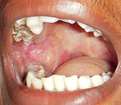
Site: In submucous fibrosis, fibrotic changes are frequently seen in the buccal mucosa, retromolar area, uvula, soft palate, palatal
fauces, tongue, lips, pharynx and esophagus, etc. It is believed, that the disease initiates from the posterior part of the oral cavity and then it gradually spreads to the anterior locations.

PRESENTATION
In the initial phases of the disease, palpation of the mucosa elicits a "wet leathery" feeling.

Petechial spots may also be seen in the early stages of the disease over the mucosal surfaces of tongue, lips and cheek, etc.

Oral mucous membrane is very painful upon palpation at this stage.

One of the most important characteristic features of oral submucous fibrosis is the gradual stiffening of the oral mucosa with progressive reduction in the mouth opening (trismus).

In mild cases, there may be white areas on the soft palate, but in severe cases, it shows restricted movements. Patients also have a 'bud-like' shrunken uvula.

Thinning and stiffening of the lips causing microchelia and presence of circumoral fibrous bands. Areas of hypo or hyper pigmentation are seen in the oral mucosa.

Loss of stippling occurs in the gingiva, and it becomes depigmented and fibrotic.

Floor of mouth becomes blanched and it gives a leathery feeling during palpation.

Palate presents several fibrous bands, which are radiating from the pterygomandibular raphe to the anterior faucial pillars.

The faucial pillars may be thick and short and the tonsils are often placed between them.

When the disease progresses to the pharynx and esophagus, it causes extreme difficulty in deglutition.

Treatment:
Stoppage of all habits, grinding and rounding of sharp cuspal edge of teeth, routine extraction of all third molars are the preliminary steps in the treatment plan. The definitive treatment of OSF includes intralesional injections of collagenase, corticosteroids and fibrinolysis,etc. Systemic administration of steroids is also done in several cases.

Biopsy is mandatory before treatment and if the dysplastic features are present in the epithelium, steroids should be avoided from the treatment schedules.

Fovea Palatine

# Fovea palatine situated in hard palate are significant as: 
A. Termination of maxillary denture
B. Opening of minor salivary gland ducts 
C. Indicates closure of mid palatine raphe 
D. Opening of greater palatine canal


The correct answer is B. Opening of minor salivary gland ducts. 

Palatine fovea
These are the orifices of common collecting ducts of minor palatine salivary glands.

The fovea palatini are two depressions that lie bilateral to the midline of the palate, at the approximate junction between the
soft and hard palate.

They denote the sites of opening of ducts of small mucous glands of the palate. They are often useful in the identification of the
vibrating line because they generally occur with in 2 mm of the vibrating line.

The hamular process, or hamulus, is a bony projection of the medial plate of the pterygoid bone and is located distal to the
maxillary tuberosity. Lying between the maxillary-tuberosity and the hamulus is a groove called the hamular notch This notch
is a key clinical landmark in maxilla) denture construction because the maximum posterior extent of the denture is the vibrating line that runs bilaterally through the hamular notches.

The hamulus can be palpated clinically and it can be a possible site of irritation in denture wearing patients, touches this process. The tendon of the tensor villi palatine muscle runs across the hamulus to reach the soft palate. Under the
tendon is a small bursa (membrane between the moving tendon and the hamulus. Inflammation and pain can result from the denture mechanical irritation by unstable dentures.

Gillies Temporal approach

# In Gillies Temporal approach for reduction of zygomatic arch fracture, Rowes elevator is placed between:  
A. Superficial facia and temporal fascia  
B. Temporal bone and temporalis muscle  
C. Temporal fascia and Temporalis muscle
D. Skin and superficial fascia


The correct answer is C. Temporal Fascia and Temporal muscle. 

In Gillies temporal fascia for reduction of zygomatic arch fracture, elevator is placed between temporal fascia and Temporalis muscle.


The temporal fascia is attached to the zygomatic arch and the temporal muscle passes downward medial to the fascia to be
attached to the coronoid process.Between these two structures a natural anatomical space exists into which an instrument can be inserted and it can be utilized to elevate the displaced zygoma on its arch into position.

Technique: The hair is shaved from the temporal region of the scalp. The external auditory meatus is plugged with cotton to
prevent any fluid or blood getting inside.
An incision about 2 to 2.5 cm in length is made, inclined forward at an angle of 45 degrees to the zygomatic arch, well in the
temporal region. Care is taken to avoid injury to the superficial temporal vessels. The temporal fascia is exposed which can be identified as white glistening structure. The incision is taken into the fascia and the fibres of temporalis muscles will be seen. Long Bristow's periosteal elevator is passed below the fascia and above the muscle.

Once this correct plane is identified and instrument is inserted through it downward and forward, the tip of
the instrument is adjusted medially to the displaced fragment.

A thick gauze pad is kept on the lateral aspect of the skull to protect it from the pressure of elevator while reduction is going on.

The operator has to grasp the handle of the elevator with both hands and assistant has to stabilize the head of the patient. (During elevation procedure care should be taken that pressure is not exerted on the lateral surface of the skull to end up with depressed fracture of the skull).

The tip of the elevator is manipulated upward, forward and outward. The snap sound will be heard as soon as reduction procedure is complete. Wound is
closed in layers after withdrawing the elevator.

Care is taken that after surgery at least for 5 to 7 days, no pressure is exerted on the area till the bone consolidates. Patient is instructed to sleep in supine position or not to sleep on the operated side.