SEARCH:

Oral Pathology Histopathological Diagrams - All in one - Histology made easy


This post contains 33 sorted diagrams of histopathological pictures of pathologies related to oral and maxillofacial regions. From benign to malignant neoplasms of oral cavity, salivary gland tumors, cyst and tumors of jaws and oral cavity, to lesions affecting nerves and muscles and soft tissues, it contains all you need to have a good command in oral pathology. 

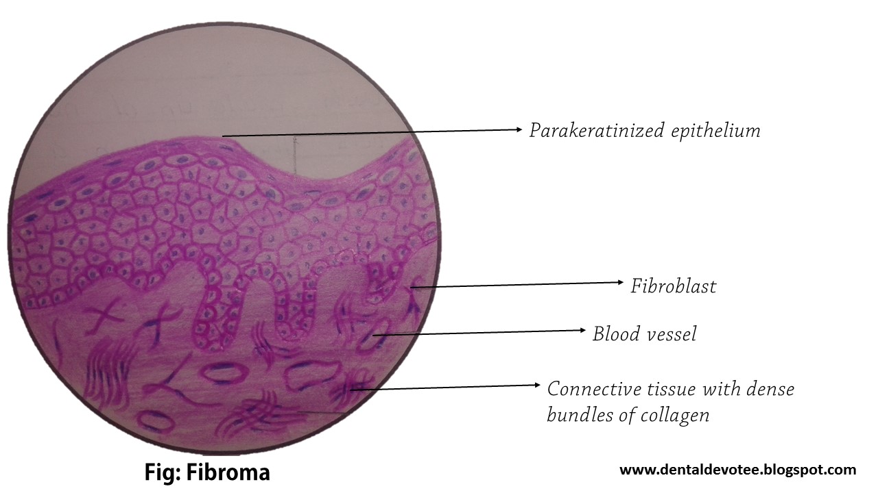
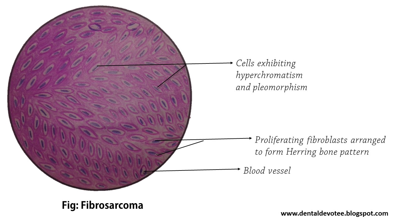
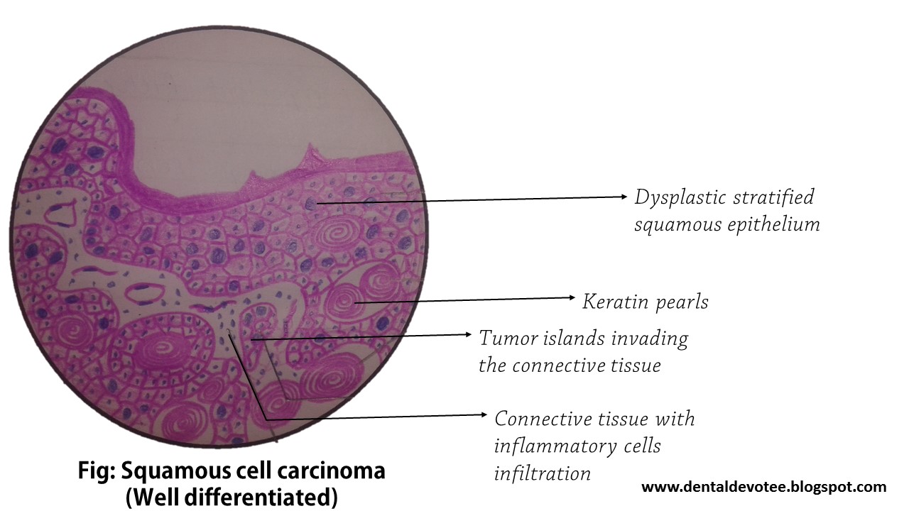
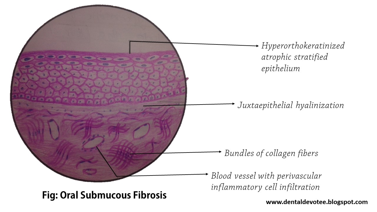
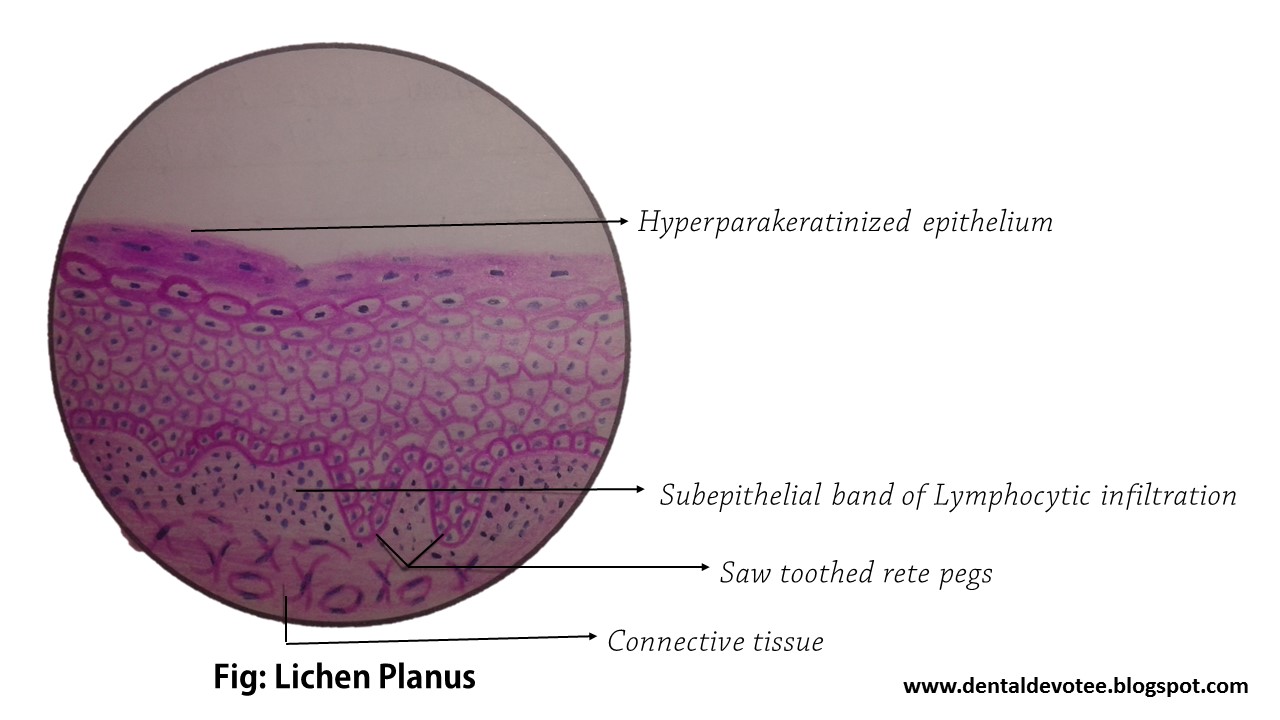
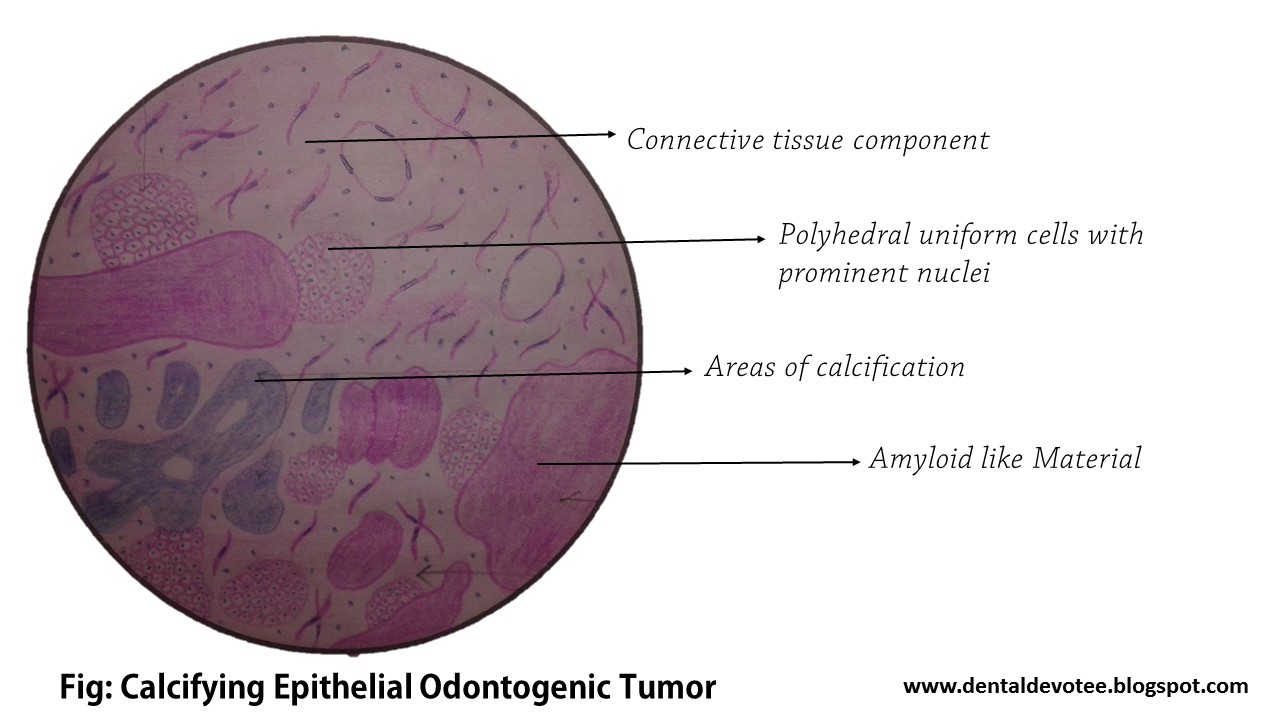
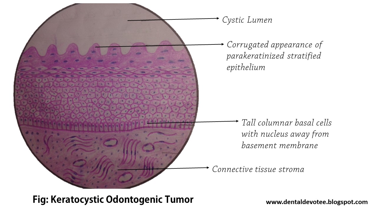
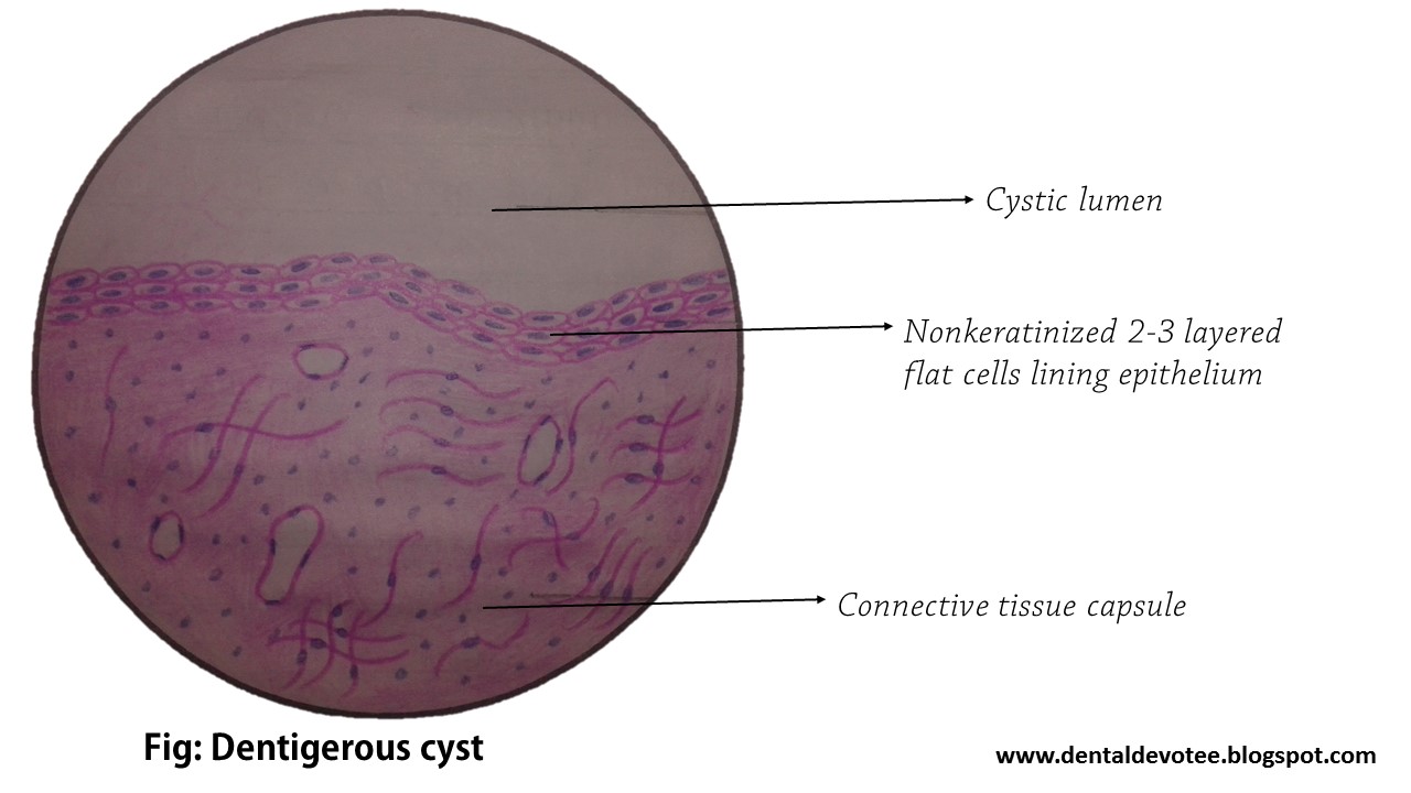
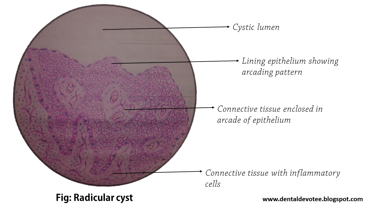
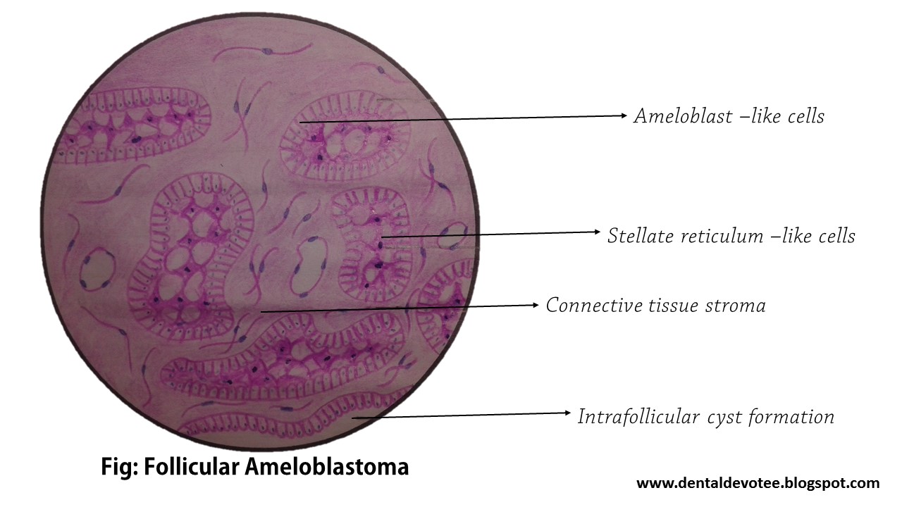
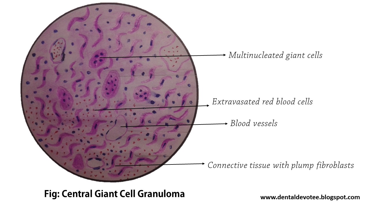
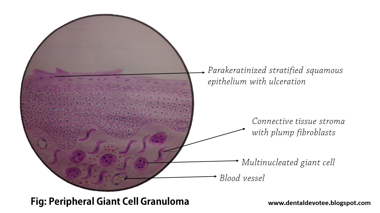
These diagrams were drawn with Hematoxylin and Eosin colored Pencils by Raman Dhungel during his Bachelor of Dental Surgery course on third year at B.P. Koirala Institute of Health Sciences, BPKIHS Dharan.  The slides included in this video are: Fibroma, FIbrosarcoma, Papilloma, Verrucous carcinoma, Mild dysplasia, Carcinoma in situ, Squamous cell carcinoma (well differentiated), Neurilemmoma, Neurofibroma, Oral submucous fibrosis, cavernous hemangioma, Lipoma, Lichen planus, malignant melanoma, osteosarcoma, pleomorphic adenoma, warthin's tumor, Adenoid cystic carcinoma, Mucoepidermoid carcinoma, Calcifying epithelial odontogenic tumor, Adenomatoid odontogenic tumor, Keratocystic odontogenic tumor, Dentigerous cyst, Radicular cyst, Plexiform ameloblastoma, Follicular ameloblastoma, Central giant cell granuloma, Peripheral giant cell granuloma, Fibrous dysplasia, Paget's disease of bone, Pemphigus, Pemphigoid (Bullous pemphigoid), and pyogenic granuloma.

Much time and effort was needed to produce this post. We would be gratified to know that if this postbecomes of some help to you. Please share with your friends and students too. 


Behcet's Syndrome

Behçet’s syndrome is a multisystem disease that predominantly affects young males and is
characterized by multiple superficial, painful “aphthous-like ulcers” in the oral cavity.
However to fulfill the criteria of being Behçet’s syndrome, clinically there should be a presence of an aphthous-like ulcer in the oral cavity along with at least two of the following lesions e.g. skin lesion, eye lesion or genital lesion, etc.

ETIOLOGY
Etiology of Behcet’s syndrome is unknown; however, the disease is believed to be caused by
some immunologic abnormality.

CLINICAL FEATURES
Oral lesions: Aphthous-like ulceration in the oral cavity.

Skin lesions: Erythematous macular, papular, vesicular or pustular lesions in the skin; thrombophlebitis may also sometimes develop.

Eye lesions: Ocular lesions in Behçet’s syndrome include uveitis, conjunctivitis, photophobia and retinitis, etc.

Genital lesions: Ulceration in the genitalia, which looks similar to those of the oral cavity.

Other lesions: Behcet’s syndrome sometimes presents some additional features like neural,
vascular, articular, renal or gastrointestinal lesions of various kinds.

HISTOPATHOLOGY
Microscopically the lesions produce similar feature to what is found in a minor aphthous ulcer. However, there can be some additional features like severe vasculitis and vascular damage, etc.

TREATMENT
Behçet’s syndrome is treated by systemic steroid therapy.

Pyogenic Granuloma

Pyogenic granuloma represents an over-exuberant tissue reaction to some known stimuli or injuries. The term pyogenic granuloma is somewhat a misnomer since the condition is not associated with pus formation.

CLINICAL FEATURES
Age: occurs at an early age.
Sex: seen more frequently in females.
Site: mostly occurs in relation to the gingiva, however on rare occasions, other mucosal sites may be involved.

PRESENTATION
• The lesion appears as a small, pedunculated or sessile, painless, soft, lobulated growth on the gingiva

• Labial surface of the gingiva is more frequently affected than the lingual surface.

• The lesion is often ulcerated and bleeds profusely, either upon provocation or spontaneously.

• The ulcerated area of the lesion is often covered by a yellow fibrinous membrane.

• The rate of growth of the lesion is very rapid and its maximum size could be up to 1 cm in
diameter

•Untreated lesion of pyogenic granuloma undergoes fibrosis due to decreased vascularity and in such cases it appears small, firm with little tendency to bleed. This lesion is called “fibroepithelial polyp”

• Similar lesions appearing on the gingival tissue of pregnant women are known as “pregnancy tumor”.

HISTOPATHOLOGY 
• Histologically, the lesion is composed of lobular masses of hyperplastic granulation tissue, containing multiple proliferating fibroblasts, many blood capillaries and a variable number of chronic inflammatory cells.

• The lesion is a vascular one and it occurs due to the proliferation of the endothelial cells.

• The overlying epithelium is thin and ulcerated, and in most of the cases the underlying
connective tissue shows intercellular edema.

• Areas of hemorrhage and hemosiderin pigmentation are often seen within the connective tissue stroma.

TREATMENT
Pyogenic granuloma is treated by surgical excision.

Oral Biology Past Question - 2nd Year BDS BPKIHS March 2016

Time: 2 Hours                             Total Marks: 125
1. Mention five points of difference between cellular and acellular cementum.    - 5

2. Mention five points of difference between primary and permanent pulp tissues.    - 5

3. Define Crest of Curvature. Describe the Crest of Curvature of a maxillary canine from all four aspects with a neat labeled diagram.    - 2+4 = 6

4. Describe six points of difference between primary and permanent human dentition.      - 6

5. Describe the labial aspect of the permanent mandibular right lateral incisor.      - 6

6. List and discuss the functions of dental pulp.    - 6

7. Mention the different types of the cementoenamel junction and discuss the formation of any one.  - 6

8. Discuss the phenomenon of formation of Hunter-Schreger band.      - 6

9. Describe late bell stage of amelogenesis with suitable diagram.       - 4+3 = 7

10. List the transitory structures formed during the development of teeth and describe any one.     - 7

11. Discuss the development of periodontal ligament fibers.     - 7

12. Discuss the formation of the root of the permanent mandibular central incisor.   - 7

13. Describe the process of modulation in amelogenesis with suitable diagrams.      - 7

14. Discuss the keyhole pattern of enamel.    - 7

15. Discuss reciprocal induction in context to the development of teeth.     -7

16.  Discuss in detail the anatomy of the permanent maxillary right central incisor.    -10


17. WRITE SHORT NOTES ON: (3*5 = 15)
a. Perikymata
b. Prismless enamel
c. Intermediate plexus
d. Neural crest cells
e. Plexus of Rashkow

18. Define the following. (1*5 = 5)
a. Fossa
b. Ridge
c. Lobe
d. Cingulum
e. Point angle