SEARCH:

Sensory nerve supply to the base of the tongue:

 # Sensory nerve supply to the base of the tongue:
A. Facial nerve
B. Trigeminal nerve
C. Glossopharyngeal nerve
D. Optic nerve


The correct answer is C. Glossopharyngeal nerve.

The hypoglossal nerve (CN XII) provides motor innervation to all of the intrinsic and extrinsic muscles of the tongue except for the palatoglossus muscle, which is innervated by the vagus nerve (CN X). It runs superficial to the hyoglossus muscle. Lesions of the hypoglossal nerve cause deviation of the tongue to the ipsilateral (i.e., damaged) side.

Taste to the anterior two-thirds of the tongue is achieved through innervation from the chorda tympani nerve, a branch of the facial nerve (CN VII). General sensation to the anterior two-thirds of the tongue is by innervation from the lingual nerve, a branch of the mandibular branch of the trigeminal nerve (CN V3). The lingual nerve is located deep and medial to the hyoglossus muscle and is associated with the submandibular ganglion.

On the other hand, taste perception in the posterior third of the tongue is accomplished through innervation from the glossopharyngeal nerve (CN IX), which also provides general sensation to the posterior one-third of the tongue. Taste perception also is performed by both the epiglottis and the epiglottic region of the tongue, which receives taste and general sensation from innervation by the internal laryngeal branch of the vagus nerve (CN X). Damage to the vagus nerve (CN X) causes contralateral deviation (away from the injured side) of the uvula.

Reference: Dotiwala AK, Samra NS. Anatomy, Head and Neck, Tongue. [Updated 2022 Aug 27]. In: StatPearls [Internet]. Treasure Island (FL): StatPearls Publishing; 2023 Jan-. Available from: https://www.ncbi.nlm.nih.gov/books/NBK507782/

Sensory nerves supply to the body of the tongue:

 # Sensory nerves supply to the body of the tongue:
A. Optic nerve
B. Trigeminal nerve
C. Facial nerve
D. Glossopharyngeal nerve


The correct answer is B. Trigeminal nerve.

The hypoglossal nerve (CN XII) provides motor innervation to all of the intrinsic and extrinsic muscles of the tongue except for the palatoglossus muscle, which is innervated by the vagus nerve (CN X). It runs superficial to the hyoglossus muscle. Lesions of the hypoglossal nerve cause deviation of the tongue to the ipsilateral (i.e., damaged) side.

Taste to the anterior two-thirds of the tongue is achieved through innervation from the chorda tympani nerve, a branch of the facial nerve (CN VII). General sensation to the anterior two-thirds of the tongue is by innervation from the lingual nerve, a branch of the mandibular branch of the trigeminal nerve (CN V3). The lingual nerve is located deep and medial to the hyoglossus muscle and is associated with the submandibular ganglion.

On the other hand, taste perception in the posterior third of the tongue is accomplished through innervation from the glossopharyngeal nerve (CN IX), which also provides general sensation to the posterior one-third of the tongue. Taste perception also is performed by both the epiglottis and the epiglottic region of the tongue, which receives taste and general sensation from innervation by the internal laryngeal branch of the vagus nerve (CN X). Damage to the vagus nerve (CN X) causes contralateral deviation (away from the injured side) of the uvula.

Reference: Dotiwala AK, Samra NS. Anatomy, Head and Neck, Tongue. [Updated 2022 Aug 27]. In: StatPearls [Internet]. Treasure Island (FL): StatPearls Publishing; 2023 Jan-. Available from: https://www.ncbi.nlm.nih.gov/books/NBK507782/

From which arch is hypobranchial eminence derived?

 # Hypobranchial eminence is derived from:
A. 1st arch 
B. 2nd arch
C. 3rd arch 
D. 4th arch


The correct answer is C. 3rd arch.

During embryonic development, the pharyngeal arches play a crucial role in the formation of various structures in the head and neck region. The third pharyngeal arch gives rise to several important structures, including the hyoid bone, the posterior one-third of the tongue (base of the tongue), and the associated muscles and nerves.

The hypobranchial eminence is a swelling within the third pharyngeal arch that contributes to the formation of the base of the tongue. It gives rise to the posterior part of the tongue, while the anterior part of the tongue is formed by the first pharyngeal arch.

Therefore, the correct option is C. 3rd arch.

Development of coronoid process of mandible occurs at:

 # Development of coronoid process of mandible occurs at:
A. 10-12 weeks 
B. 12-16 weeks
C. 10-14 weeks 
D. 14-16 weeks


The correct answer is C. 10-14 weeks.

The development of the coronoid process of the mandible occurs during the embryonic period of gestation. Specifically, it takes place between 10 and 14 weeks of gestation. Therefore, the correct option is C. 10-14 weeks.

During this time frame, the mandible undergoes various developmental processes, including the formation of the mandibular body, the ramus, and the coronoid process. The coronoid process is a bony prominence located on the anterior aspect of the ramus of the mandible. It serves as an attachment site for muscles involved in chewing and jaw movement.

Options A and B (10-12 weeks and 12-16 weeks) and Option D (14-16 weeks) do not accurately represent the developmental period when the coronoid process of the mandible forms. The correct time frame is 10-14 weeks.

Posterior one third of the tongue or base is formed by:

 # Posterior one third of the tongue or base is formed by:
A. Two lingual swelling
B. Hypobranchial eminence
C. Copula
D. Tuberculum impar


The correct answer is B. Hypobranchial eminence.

The hypobranchial eminence is a swelling in the embryonic development of the tongue that gives rise to the base of the tongue. It is located in the midline and forms the posterior part of the tongue. It develops from the third and fourth pharyngeal arches during embryonic development.

The other options mentioned are not specifically related to the formation of the posterior one third of the tongue.

A. Two lingual swellings: These refer to the anterior two-thirds of the tongue, which are formed by a pair of lingual swellings that merge during development.

C. Copula: The copula is a structure in embryonic development that contributes to the formation of the posterior part of the tongue, but it does not specifically form the base of the tongue.

D. Tuberculum impar: The tuberculum impar is a small median swelling in the embryonic development of the tongue, located at the midline in the anterior part of the tongue, and it does not contribute to the formation of the base of the tongue.

Therefore, the correct option is B. Hypobranchial eminence.

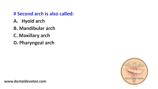
Second arch is also called:

 # Second arch is also called:
A. Hyoid arch 
B. Mandibular arch
C. Maxillary arch 
D. Pharyngeal arch


The correct answer is A. Hyoid arch. 

The second arch in embryonic development is indeed referred to as the Hyoid arch. The hyoid arch, also known as the second pharyngeal arch, is one of the series of pharyngeal arches that contribute to the development of structures in the head and neck.

The hyoid arch gives rise to important structures such as the lower part of the hyoid bone (a bone in the neck), the muscles of facial expression, muscles involved in swallowing, and certain muscles in the throat. Nerves and blood vessels associated with this arch also develop within it.

Each pharyngeal arch has its specific developmental contributions and plays a role in the formation of different structures. The hyoid arch, or second arch, is significant in the development of various structures in the head and neck region during embryonic development.

First arch is also called:

 # First arch is also called:
A. Hyoid arch 
B. Mandibular arch
C. Maxillary arch 
D. Pharyngeal arch


The correct answer is B. Mandibular arch.

The first arch in embryonic development is often referred to as the Mandibular arch. During the early stages of embryonic development, several structures form in a series of arches known as pharyngeal arches. These arches play a crucial role in the development of the face and neck.

The Mandibular arch, also known as the first pharyngeal arch, is the most anterior of the pharyngeal arches. It gives rise to various structures including the lower jaw (mandible), certain muscles of mastication (chewing), and some muscles of facial expression. The nerves and blood vessels associated with this arch also develop within it.

Each pharyngeal arch has a specific set of components that contribute to the formation of different structures. In the case of the Mandibular arch, it primarily gives rise to the lower jaw, muscles involved in chewing, and some facial muscles. The arches undergo complex growth, fusion, and differentiation processes to form the various structures of the face and neck during embryonic development.